Linyu Zeng1, Weiran Ling1, Hai Xiong*
1 These authors contributed equally to this work.
On May 13th, Prof. Xiong group from the Institute for Advanced Studies at Shenzhen University published a research paper titled “pH-regulated self-assembly and Ag+-responsive bioimaging of endoplasmic reticulum-targeted probe in vitro/in vivo” on Chinese Chemical Letters (https://doi.org/10.1016/j.cclet.2026.112941). Prof. Hai Xiong is the only corresponding author, and Ms. Linyu Zeng (master’s student) and Weiran Ling (undergraduate) is the first co-author. Shenzhen University is the only corresponding affiliation.
Amphipathic TAP-dU and TAP-dC were designed with a distinctive donor-π-acceptor (D-π-A) electronic architecture, which the nucleoside function and TAP-moiety act as the electron donor and electron acceptor, respectively. The base-triggered charge transfer is influenced by protonation/deprotonation states and conformational twisting. Hydrogen-bonding interactions between the pyrimidine and ribose were thoroughly examined by nuclear magnetic resonance (1H NMR) spectroscopy, single-crystal X-ray crystallography, and density functional theory (DFT) calculations. Importantly, TAP-dU exhibits pH-dependent self-assembly and π-π stacking interactions, leading to an aggregated fluorescence multicolor shift from blue to green to yellow, with emission wavelengths reaching up to 545 nm (Fig.1).
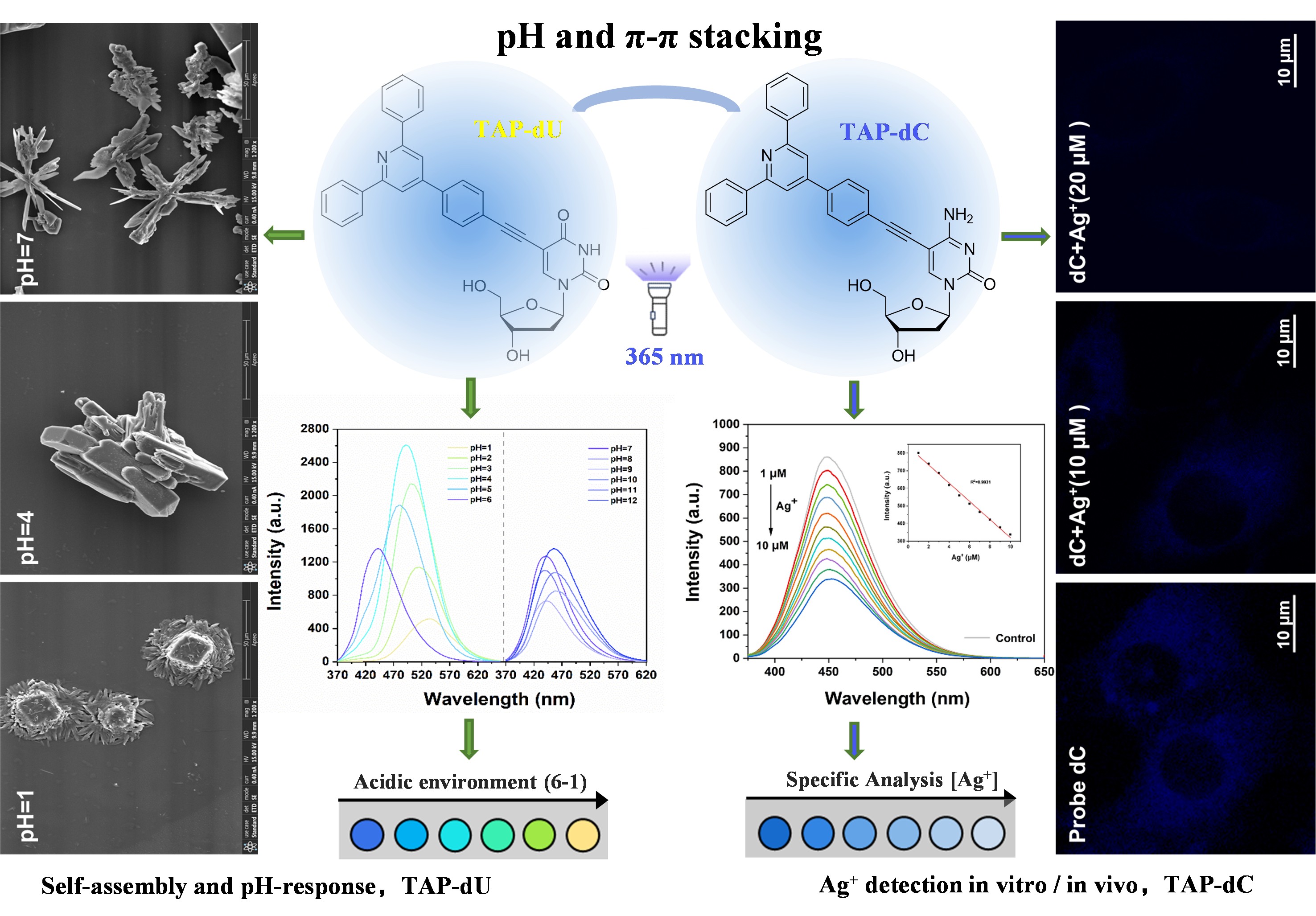
Fig. 1. Endoplasmic reticulum-targeted TAP-dU exhibits pH-dependent self-assembly and TAP-dC serves as a promising candidate for Ag+-responsive bioimaging in vitro/in vivo.
Unlike our previously reported nucleus-targeted TPE-dU/TPE-dC and mitochondria-targeted TPE-FdU (Chinese Chemical Letters 2021, 32, 1687–1690; Analytical Chemistry 2023, 95, 18880−18888), this study demonstrates that both nontoxic TAP-dC and TAP-dU effectively achieve endoplasmic reticulum (ER)-specific imaging in NIH-3T3 and HeLa cells. Additionally, due to its biocompatibility, TAP-dC also serves as a promising candidate for in vivo Ag+ detection in zebrafish via bioimaging (Fig.2).
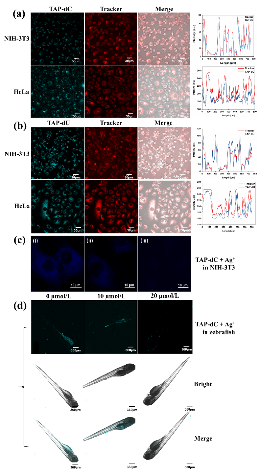
Fig. 2. (a) CLSM images of NIH-3T3 and HeLa cells after coincubation with TPA-dC (20 μmol/L) at 4 h. (b) after coincubation with TPA-dU (20 μmol/L) at 4 h. Fluorescence imaging of Ag+ concentration-dependent incubated with TAP-dC at pH 7 for 1h (i) 0 μmol/L, (ii) 10 μmol/L, (iii) 20 μmol/L, (c) in NIH-3T3 and (d) in zebrafish. λex = 800 nm, λem = 500–550 nm. Scale bar: 360 mm.



